Before you book
Share the exact test name on your prescription with our team. We will confirm the appointment slot, final price, preparation instructions and expected report availability for your test.
Please tell the team about any assistance you need for your visit. Bring your prescription and previous reports if available.
Visit our Dilshad Garden centre or call +91 99531 43636 for booking information.
Complete echocardiography — 2D Echo, Colour Doppler, M-Mode and Tissue Doppler. Assessment of heart function, valve disease and ejection fraction. Female radiologist Dr Samta Parteki MD & Dr Saloni Jain MD available. Same-day reports on WhatsApp.
A Live Ultrasound of Your Beating Heart
A 2D Echo (Two-Dimensional Echocardiography) is an ultrasound scan of the heart. Using high-frequency sound waves, it produces real-time moving images of the heart's chambers, valves, walls and major blood vessels — giving your cardiologist a live picture of how your heart is functioning.
Unlike an ECG, which records only the heart's electrical activity, a 2D Echo shows the heart's actual physical structure and mechanical movement — how strongly the heart muscle contracts, whether valves open and close correctly, and whether any fluid surrounds the heart.
It is completely painless, non-invasive, uses no radiation, and requires no preparation. The test takes 20–30 minutes and reports are delivered the same day via WhatsApp.
⏰ Mon–Sun · 8:00 AM – 8:00 PM
🚇 5 min from Dilshad Garden Metro (Red Line)
Types of Echocardiography at Adhiraj Imaging
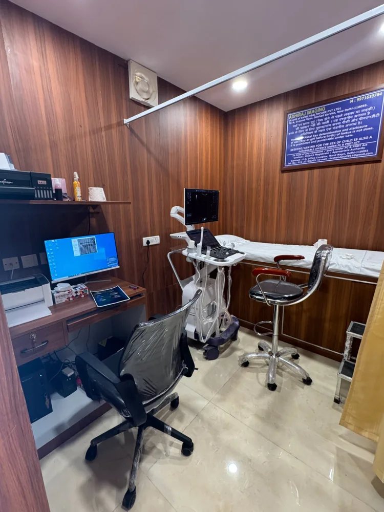
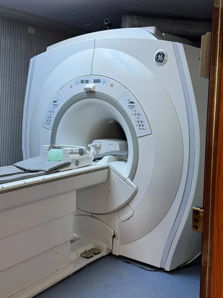
All echo modalities performed on a high-resolution GE cardiac ultrasound system. Reports by MD radiologist — same day.
2D Echocardiography
The core echo study. Real-time two-dimensional images of all four heart chambers, walls and valves. Measures ejection fraction (EF), wall thickness, and overall systolic function. Recommended as the first-line cardiac imaging test.
Colour Doppler Echo
Colour Doppler is added to 2D Echo to map blood flow direction and velocity through each valve. Essential for detecting valve leaks (regurgitation), narrowing (stenosis), septal defects and pulmonary hypertension.
M-Mode Echocardiography
A single ultrasound beam records heart structure movement over time. The most accurate method for measuring chamber dimensions, wall thickness and valve motion — used for precise LV size quantification.
Tissue Doppler Imaging (TDI)
Measures velocity of the heart muscle itself. TDI is the most sensitive method for detecting diastolic dysfunction — often the earliest sign of heart disease in hypertension and diabetes, missed by conventional echo.
When is a 2D Echo Recommended?
A 2D Echo is one of the most commonly requested cardiac investigations. Here are the most frequent reasons doctors prescribe it.
Chest Pain & Breathlessness
Rules out or confirms cardiac cause. Identifies wall motion abnormalities, pericardial effusion and valve disease.
Heart Failure
Measures EF, identifies dilated or hypertrophic cardiomyopathy. Essential for diagnosing and grading heart failure severity.
Hypertension
Assesses LV hypertrophy and diastolic dysfunction — the early cardiac effects of long-standing high blood pressure.
Valve Disease
Diagnoses and grades mitral stenosis, mitral regurgitation, aortic stenosis, aortic regurgitation and tricuspid disease.
After Heart Attack
Evaluates post-MI wall motion, left ventricular function and complications such as VSD or pericardial effusion.
Pre-operative Evaluation
Mandatory before cardiac surgery. Also done before major non-cardiac surgery in patients with known or suspected heart disease.
Palpitations
Identifies structural causes of palpitations — enlarged chambers, hypertrophic cardiomyopathy — that may predispose to arrhythmias.
Diabetes Cardiac Screening
Screens for diabetic cardiomyopathy and silent LV dysfunction — common in diabetic patients with no cardiac symptoms.
Congenital Heart Disease
Diagnoses ASD, VSD, PDA and other structural defects in children and adults. Colour Doppler is essential for shunt detection.
What Your 2D Echo Report Covers
A standard 2D Echo with Colour Doppler at Adhiraj Imaging covers all the parameters below — reported by our MD radiologist on the same day.
Ejection Fraction (EF)
% of blood pumped per beat. Normal: 55–70%. EF below 40% indicates significant heart failure.
Chamber Size (LV, RV, LA, RA)
Enlarged chambers indicate pressure or volume overload from valve disease or cardiomyopathy.
Wall Thickness & Motion
LV hypertrophy from hypertension, thinning from old infarct, regional wall motion abnormalities (RWMA).
E/A Ratio & TDI
Diastolic dysfunction graded 1–3. Often the earliest sign of heart disease in hypertension and diabetes.
Mitral & Aortic Valves
Valve area, pressure gradient, regurgitation severity graded mild / moderate / severe. Guides surgical decisions.
Pulmonary Artery Pressure
PASP estimated from TR jet. Elevated PASP (>35 mmHg) indicates pulmonary hypertension.
Pericardial Effusion
Fluid around the heart quantified as trivial / mild / moderate / large. Large effusion may need drainage.
Aortic Root & IVC
Aortic root dilatation screened. IVC size and collapsibility estimates right atrial pressure.
Colour Doppler Flow
Colour-coded maps of blood flow through all valves — instantly shows regurgitation jets and abnormal shunts.
2D Echo Near You
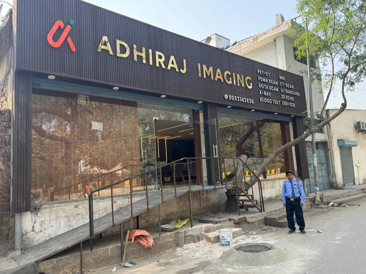
Adhiraj Imaging is in Dilshad Garden, East Delhi — easily reachable from all nearby areas by metro, auto or car within 20 minutes.
76-A, GF1, Opp. IHBAS Hospital Gate No. 3
Direct auto from Shahdara Metro Station (Red Line)
Via NH-9 or Vivek Vihar flyover
Via GTB Nagar or Dilshad Colony road
Via Vikas Marg, easily accessible
Metro interchange station — Red Line direct
Loni, Mohan Nagar, Sahibabad via NH-9
Sector 62/63 via metro interchange
Before Your 2D Echo — What to Expect
The good news — a 2D Echo requires almost no preparation. Here is exactly what to expect.
No Fasting Required
You can eat and drink normally. No fasting is needed for a standard 2D Echo. Simply walk in with your doctor's prescription during our working hours.
✓ Walk-in welcome any time 8AM–8PMWear a Loose Top or Shirt
Wear clothing that can be easily opened at the front. A small amount of gel is applied to the chest — it is warm, painless and wipes off easily.
Bring Your Doctor's Prescription
A valid prescription from your cardiologist, physician or GP is required. Bring any previous echo or ECG reports if available — this helps our radiologist provide context-aware reporting.
⚠ Prescription mandatory for all cardiac testsThe Test Takes 20–30 Minutes
You lie on your left side on the exam table. The sonographer moves a small probe across your chest in different positions. You will see live images of your heart on the screen. Completely painless.
Report Ready Same Day
Your 2D Echo report is reviewed by our MD radiologist and sent via WhatsApp and email the same day. Hard copy available at the centre. Urgent reports can be done in 1–2 hours.
✓ Report via WhatsApp · Hard copy availableWhatsApp or call — we'll confirm your slot and send a reminder.
⏰ Mon–Sun · 8:00 AM – 8:00 PM
🚇 Dilshad Garden Metro · Red Line · 5 min walk
Why Patients Choose Adhiraj Imaging for 2D Echo
Trusted by patients across East Delhi, Shahdara, Vivek Vihar, Krishna Nagar and Ghaziabad at our Dilshad Garden centre.
Common Questions About 2D Echo
Answers to questions we hear most before the scan.
Is a 2D Echo the same as a heart ultrasound?
Do I need to fast before a 2D Echo?
What is a normal ejection fraction on 2D Echo?
What is the difference between a 2D Echo and Colour Doppler Echo?
Is a female radiologist available for 2D Echo?
When will I receive my 2D Echo report?
Is 2D Echo covered under CGHS or insurance?
Book Your 2D Echo Today
No fasting · Walk-in welcome · Reports in 1–2 hours · Female radiologist available · East Delhi